Biomarkers/diagnostics/blood tests thread

Plasma proteome profiling of healthy individuals across the life span in a Sicilian cohort with long-lived individuals https://onlinelibrary.wiley.com/doi/10.1111/acel.13684

Confirmed correlation 2/ age of fibulin-1, dystroglycan, gamma-glutamyl hydrolase, & ID’d novel proteins correlating w/ age

@AgingCell

https://onlinelibrary.wiley.com/doi/10.1111/psyp.14113

Older adults had lower (slower) alpha center frequencies than younger adults (younger = 10.7 Hz, older = 9.6 Hz; t 28 = 2.20, P = 0.036; Cohen’s d = 0.79) and lower aperiodic-adjusted alpha power (younger = 0.78 μV2, older = 0.45 μV2; t 28 = 2.52, P = 0.018; Cohen’s d = 0.93), although bandwidth did not differ between groups (younger = 1.9 Hz, older = 1.8 Hz; t 28 = 0.48, P = 0.632; Cohen’s d = 0.17) (Fig. 5c). The mean aperiodic-adjusted alpha power difference between groups was 0.33 μV2 Hz−1, whereas, when comparing total (nonaperiodic-adjusted) alpha power, the mean difference was 0.45 μV2 Hz−1. This demonstrates that, although alpha power changes with age, the magnitude of this change is exaggerated by conflating age-related alpha changes with age-related aperiodic changes.

Regarding aperiodic activity, older adults had lower aperiodic offsets (younger = −11.1 μV2, older = −11.9 μV2; t 28 = 6.75, P < 0.0001; Cohen’s d = 2.45) and lower (flatter) aperiodic exponents (younger = 1.43 μV2 Hz−1, older = 0.75 μV2 Hz−1; t 28 = 7.19, P < 0.0001; Cohen’s d = 2.63) (Fig. 5e). Participant-specific aperiodic components were reconstructed based on individual offset and exponent parameter fits from channel Cz, and used to compare frequency-by-frequency differences between groups (Fig. 5d). From reconstructions, significant differences were found between groups in the frequency ranges 1.0–10.5 Hz and 40.2–45.0 Hz (P < 0.05, uncorrected t-tests at each frequency). This demonstrates, in real data, how group differences in what would traditionally be considered to be oscillatory bands can actually be caused by aperiodic—nonoscillatory—differences between groups (compare with Fig. 1).

Uploads


50:31 Now playing

EEG2

2 views52 minutes ago


32:19 Now playing

EEG

3 views1 hour ago

age and neurologic disease increase the noise of the systemic proteome, most noticeably when looking at uPAR, TRAIL R1, IL-16, TIMP-1, IL-15R alpha, CD27, and APJ, TNFRSF27, CCL25, and TGFBR2

To test if the 10 protein biomarkers would enable a direct quantification of person’s biological age, we calculated the mean of their SDs (10-protein noise) and plotted these values vs. chronological age of the young (20–30 years), middle-aged (50–60), and old (70 +) individuals

in GrimAge https://www.ncbi.nlm.nih.gov/pmc/articles/PMC6366976/

However, age-adjusted DNAm PAI-1 outperforms AgeAccelGrim for several age-related traits

Markers of inflammation and metabolic conditions are associated with several epigenetic biomarkers including AgeAccelGrim, age-adjusted DNAm TIMP-1, and DNAm PAI-1. However, DNAm PAI-1 stands out when it comes to associations with type 2 diabetes status, glucose-, insulin-, triglyceride levels, anthropometric measures of adiposity (body mass index and waist-to-hip ratio), and computed tomography data on fatty liver and excess adipose tissue.

Our DNAm-based surrogate biomarkers of plasma protein levels may be leveraged by researchers who rely on bio-banked DNA samples without the availability of plasma samples. Strong evidence supports links between plasma proteins used in the construction of GrimAge and various age-related conditions: ADM levels are increased in individuals with hypertension and heart failure [41]. Plasma B2M is a clinical biomarker associated with cardiovascular disease, kidney function, and inflammation [42]. Plasma cystatin-C is used to assess kidney function [43]. ADM, B2M, cystatin C, and leptin relate to many age-related traits including cognitive functioning [4446]. GDF-15 is involved in age-related mitochondrial dysfunction [46]. PAI-1 plays a central role in a number of age-related subclinical and clinical conditions [47], and recent genetic studies link PAI-1 to lifespan [48]. The tissue inhibitor of metalloproteinases, TIMP-1, plays an anti-apoptotic function [49]. We acknowledge the following limitations. The levels of relatively few plasma proteins (12 out of 88) were accurately imputed based on DNAm levels in blood. In the FHS data, the measurement of the plasma proteins (exam 7) preceded the measurement of blood DNAm data (exam 8) by 6.6 years, suggesting that the DNAm profiles may not represent a highly accurate snapshot of the status of these proteins at the time of blood collection. That said, the elucidation of cause-and-effect relationships between plasma proteins and DNAm will require future longitudinal cohort studies and mechanistic evaluations.

Young Blood for Old Brains with Tony Wyss-Coray - YouTube [more important]

6:37: a 70 year old w/biological age of 35?!??!?!? is this omics/better than epigenetic? what clock used? 5:33: measure > 100 hormone-like proteins in 295 human blood samples (20-89 years) to discover protein signatures of aging [but I don’t think this is the most diagnostic, seems to be growth factor based]

CCL11 is inflammation biomarker lower in centenarians

To further elucidate the temporal relation between EBV infection and MS, we measured serum concentrations of neurofilament light chain (sNfL), a sensitive, albeit not disease-specific, biomarker of ongoing neuroaxonal degeneration (18), using an ultrasensitive single-molecule assay (19) in the samples from those who were EBV-negative at baseline. We have previously reported that sNfL levels increase as early as 6 years before clinical MS onset and may be a more accurate marker of the time of initiation of the disease process (20).

Researchers developed an algorithm called QUARTZ, based on retinal images from tens of thousands of adults between 40 and 69 years-old. It focused on the width, area, and curvature (or tortuosity) of tiny blood vessels called arterioles and venules. The team compared the performance of QUARTZ with the widely used Framingham Risk Scores framework – both separately and jointly.

https://twc-stanford.shinyapps.io/aging_plasma_proteome/ (ECM proteins change A LOT w/aging) → does glucosamine reduce it? https://www.nature.com/articles/s41591-019-0673-2 LOOK at the supplementary data it’s more useful than the shinyapps

https://www.science.org/doi/10.1126/sciadv.add6155 => increases in unsynthesized precursors like free amino acids AND 5-hydroxytryptophan, which is a precursor to serotonin

centenarians had higher phenylalanine (anti-inflammatory), higher sphingomyelins, and lower glycerophosphocholine (related to senescent cells)

metabolites positively associated with aging include purine degradation compounds (26), complement protein peptides (26), ornithine (41), PUFAs (45), metabolites related to the cytochrome P450 system (51), and kynurenic acid (55). Metabolites that were negatively associated with aging include glutamate (50), although other studies have identified an increase in glutamate with age (26, 31, 32). Among presumably healthy-aging centenarians, studies have found changes in tryptophan (decreased) (33), lysophosphocholines (decreased) (33), eicosanoids (33), 2-hydroxybenzoate (increased) (33), phenylalanine (increased) (35), and acetylglycoproteins (increased) (35) in centenarians relative to other elderly or younger individuals.

Oxidative stress has been another major theme in aging metabolomics (26, 30, 33, 36, 39, 41, 46, 50, 54, 55). Some of the more consistently associated metabolites are carnitines, particularly acylcarnitines (30, 36, 43, 57). Acylcarnitines tend to decrease at older ages, which may be related to the use of the carnitine-acylcarnitine shuttle in mitochondria to help mitigate oxidative stress. Carnitines more generally have been associated with age (46, 49, 52), with some studies reporting higher carnitine levels (41, 53, 54) and others lower levels (25, 36, 43). Glutathione, glutathione disulfide, ophthalmic acid (an analog of glutathione), and the glutathione/oxiglutathione ratio have also been found to decrease with age (39, 52, 55), suggesting that these antioxidant mechanisms may be compromised at older ages. Studies have also reported altered sphingolipid levels (35, 36, 42, 43), particularly an increase in sphingomyelins (30, 34), which might reflect processes related to the conversion of sphingomyelins to ceramides in relation to oxidative stress and inflammation. Other age-associated metabolites that have been interpreted as evidence of oxidative stress include carnosine and its precursor histidine (decreased with age) (30, 39, 52), eicosanoids (33), vitamin E (46), serine [both decreased (50) and increased (41)], glutamate (decreased) (50), and certain TCA cycle intermediates (increased) (54).

The age-metabolite associations between the populations had considerable overlap, including similar age associations noted for phenylacetylglutamine, 4-cresyl sulfate, HMB, and creatine

Ascorbate Quenches Singlet Oxygen/Fenton Reaction-Induced UPE in Skin

Ultraweak photon emissions (UPE) from our skin is a reliable indicator in the formation of reactive oxygen species such as singlet oxygen and/or hydroxyl radicals generated from Fenton reactions. When sodium ascorbate was added to skin biopsy prior to application of Fenton’s reagent that induces the toxic Fenton reaction, it was discovered that ultraweak photon emission was significantly suppressed due to the effective quenching of singlet oxygen and other cytotoxic free radicals [69]

from Estep’s Mindspan

Research shows that three biomarkers predict longevity in men: core body temperature (lower is better), blood levels of insulin (lower is better), and DHEA-S, the sulfated form of the sex hormone dehydroepiandrosterone (slower decline of blood levels in old age is better). The authors of this study note that these biomarkers are also associated with a calorie restriction diet in lab animals, which extends longevity relative to animals given unlimited food.

==

https://youtu.be/SQtuYnGsiCI?t=1119 (gait variability)

also see Bryan Johnson’s blueprint youtube video and his site


See profile for Karl R. Pfleger

[

Karl R. Pfleger 1st degree connection 1st Longevity Investor & Philanthropist. Founder, AgingBiotech.info.

](https://www.linkedin.com/in/karl-r-pfleger/)

11h

Nice article. Will help get wider exposure to the field in the same way Bryan Johnson’s press has. But when are you going to expand to better methylation clocks than just TruMe? The field will soon need an independent leaderboard with the best clocks plus functional measures. Unlikely that this is true LEV.

5 Replies 5 Replies on Karl R. Pfleger’s comment

[

Tom WeldonView Tom Weldon’s profile Author Founder, Chairman and CEO at Ponce De Leon Health

](https://www.linkedin.com/in/ACoAACXfJoMBf_fJbhrMa__1pY2240DVOLf5zqk)

10h

Hi Karl,
Although this regression line is impressive, 6 data points would be reasonable to forecast out two more points, obviously not forever. But the story will hopefully get exposure to the industry, which is critically important, especially now. In terms of the TruMe test, it actually has been validated against one of the later Horvath clocks, and of course, “accuracy” is simply conceptual. The biological age produced by any algorithm is simply an estimation, and whether one clock gives a different answer that is 5 years higher or lower than another, is probably not the most important point. Taking multiple readings over several years is what matters. Are the data consistent, is there a trend? I have over 4 years worth of data, and the first 3.5 years of data have an upward trend, similar to the increase in my chronologic age, then the trend is pronouncedly downward. Having worked in the Medtech field most of my life, I am very familiar with what the FDA will probably require for a new endpoint to be valid for FDA approval purposes. It will require a drug like trial run until all subjects have expired to prove that a particular clock is “accurate”. No one will do such a trial, which is why I chose compounds that are not drugs.

[

Hannah WentView Hannah Went’s profile (She/Her) • 1st Director of Operations at TruDiagnostic

](https://www.linkedin.com/in/ACoAADAEU_0B5PF2DAIsXYcTqxOUwE3KnJg7GqM)

9h (edited)

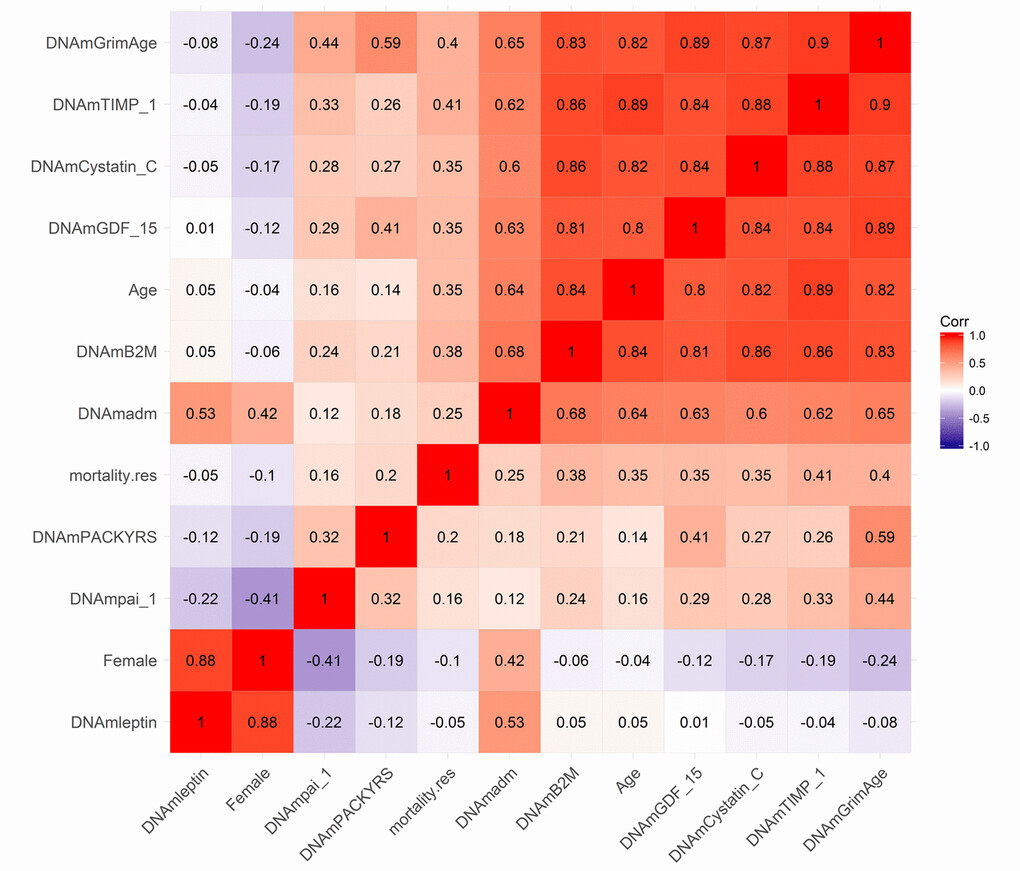
Karl R. Pfleger Tom Weldon I think another important point is that we do not want these clocks to be accurate, as that would just mean they are getting closer and closer to your chronological age - the first generation clocks. Examples of the first generation clocks include the Horvath clock, Hannum clock, etc. These clocks still have great application. For example, they were first used to help refugees seek asylum (https://www.researchgate.net/publication/327420654_European_scientists_seek_‘epigenetic_clock’_to_determine_age_of_refugees) and to identify the age of DNA at crime scenes. However, there are better predictors of biological age that are more clinically relevant - second generation clocks (and third generation - DunedinPACE). The second gen clocks are trained using an underlying aging phenotype rather than just chronological age. In addition, these clocks are much more precise than the first gen clocks, which is exactly what you would want for a clinical trial. We need to know that the results are a true change of underlying biology rather than just noise from the algorithm itself. Morgan Levine’s Principal-Component Clocks (second generation) greatly help with the precision as well!

[

Karl R. PflegerView Karl R. Pfleger’s profile • 1st Longevity Investor & Philanthropist. Founder, AgingBiotech.info.

](https://www.linkedin.com/in/ACoAACXrfxEBV4Mx4ibKNRbvWtTtsCO6XMZSm74)

8h

Hannah Went When I said better I meant in terms of mortality prediction & by extension prediction of age-related diseases. PC-GrimAge is probably the best current clock for this in research settings. Elysium has it’s Index clock (which Levine helped design when she was consulting there and which does test many sites to get the same PC benefit)—would be nice to see some good direct comparison of these 2.

DunedinPACE is also supposed to be good but the rate-of-aging measure is a different kind of thing and not as useful (IMO) when used to sample sparsely & longitudinally as Tom is doing here. (I think TruDiagnostic’s DTC clock does give a point-estimate of current bio-age too, but I don’t know the details of how that works. Note: TruDiagnostic should not be confused with TruMe labs.)

[

Karl R. PflegerView Karl R. Pfleger’s profile • 1st Longevity Investor & Philanthropist. Founder, AgingBiotech.info.

](https://www.linkedin.com/in/ACoAACXrfxEBV4Mx4ibKNRbvWtTtsCO6XMZSm74)

7h

Karl R. Pfleger Hannah, perhaps you can elaborate on your point-estimate?


See profile for Hannah Went

[

Hannah Went (She/Her) 1st degree connection 1st Director of Operations at TruDiagnostic

](https://www.linkedin.com/in/thehannahwent/)

6h

Karl R. Pfleger I agree!

As for the current clocks TruDiagnostic offers, all of them are principal component controlled. This was a large platform updated we did in spring of 2021.

As we realize the need for better predictors, TruDiagnostic is currently creating an algorithm (called OmicAge) with Harvard which was trained on 5,000 patients with full metabolomic data, large scale proteomic data, methylation and 75 clinical variables taken at 4 time points. We’ve spent the last 2.5 years creating it and will be releasing a preprint on it soon. In the meantime, I’m happy to privately send you the hazard ratio relationship to disease versus the other clocks. It significantly outperforms every clock including the DunedinPace!

Researchers at Karolinska Institutet have now shown that the level of a certain glycan structure in blood, denoted bisected N-acetylglucosamine, can be used to predict the risk of developing Alzheimer’s disease.

Went through the first 130 pages, and really it is the second page that teflon-pilled me.

It says pfas bind strongly to protein, and was used to precipitate proteins from solution as a standard laboratory practice. Later, a 3M manual to understanding pfas describes surfactants and hydrophobic/philic surface interfaces.

What I gather, is pfas are binding to all the proteins in the body and causing random noise with all protein interactions in the body.

The mouse studies do say, certain amounts of pfas exposure cause liver inflammation. Then later there are a bunch of human cancer correlations.

If PFAS are causing signaling noise and binding interference in biological systems via protein hydrophobic/philic pocket binding, and PFAS accumulate in the body over time, this is an enormous health risk that does not need a proven mechanism to regard as serious. It’s only a question of the scale of effect.

Which, albeit is always the concern with toxic pollutants. As in, what is a person or a society getting out of this vs the cost of pollution?

In the absence of highly inflationary banking I like to imagine that we could clean up and control our pollution like we handle sanitation.

AD-detect blood test… ($399)…

https://www.pnas.org/doi/10.1073/pnas.012579499

https://www.science.org/doi/10.1126/scitranslmed.abo1557
blood base biomarkers for mito dna dx damage (parkinson’s), would be very useful for testing response to amphetamines

Note the lifespan of red blood cells is precisely measured by Levitt’s
exhaled CO test (ranging from about 10 to 200 days in adults with average
around 120)

The lower your exhaled CO, the longer the lifespan of your RBC.

Expression of Most Retrotransposons in Human Blood Correlates with Biological Aging

(April 17, 2024) https://doi.org/10.1101/2024.02.09.579582

Retrotransposons (RTEs) have been postulated to reactivate with age and contribute to aging through activated innate immune response and inflammation.

Here, we systematically analyzed the relationship between RTEs expression and aging using published transcriptomic and methylomic datasets of human blood. Despite no observed correlation between RTEs activity and chronological age, most RTE classes and families except short interspersed nuclear elements (SINEs) correlate with age-associated gene signature scores. Strikingly, we found that the expression of SINEs is linked to upregulated DNA repair pathways in multiple cohorts. DNA hypomethylation with aging was observed across RTE classes and associated with increased RTEs expression in most RTE classes and families except SINEs. Additionally, our single-cell transcriptomic analysis suggests a role for plasma cells in aging mediated by RTEs. Altogether, our multi-omics analysis of large human cohorts highlights the role of RTEs in biological aging and suggests possible mechanisms and cell populations for future investigations.