Naixin Qian - Google Scholar THIS is super-relevant (esp nanoplastics but also…)
oxford instruments has A LOT of good content I’m still trying to go thru
I also visited some of the Koch Institute scanning facilities lately…
some webinar:
https://register.gotowebinar.com/recording/recordingView?webinarKey=4767004550341430543
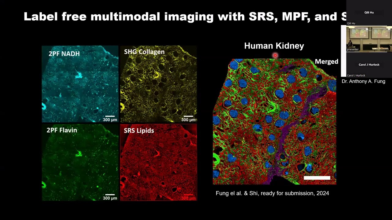
far far out but worth seeing just for the hell of it
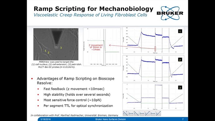
Manipulation and spectroscopy. The ability to mechan-
ically manipulate biological samples by the AFM tip
has raised the issues of measuring the forces interacting
between the AFM tip and sample and those interact-
ing between biological samples280. Force–distance curves
can be used to quantify the hydrophobic, hydrophilic,
electrostatic, van der Waals, Young’s modulus, energy
dissipation and many other physical properties of the
sample surface, provided that the properties of the AFM
tip have been defined by, for example, characteriz-
ing a reference sample. Sometimes, the tip is replaced
by a micrometre-sized bead to probe the mechanical
properties of larger surface areas of cells or tissues281.
Examples encompass measuring the elastic properties
of the cell cortex, neuronal tissues or even organs of liv-
ing animals34. However, to properly analyse the elastic
properties of a soft heterogeneous sample indented by
a sharp tip requires the application of theoretical mod-
els such as Hertz theory or similar models, which have
limitations and must be applied with care34,282. One way
to circumvent such limitations is to use a bare AFM
cantilever without the tip in a parallel plate assay, which
has shown that mammalian cells measure confining dis-
tances of their surrounding environment283 and round
up for mitosis by generating hydrostatic pressure284,285.
The physical properties, measured as force–distance
or force–time curves, depend on the speed or time at
which they are probed by AFM34. Consequently, the
elastic properties of biological systems probed at various
speeds can differ considerably286,287. To properly describe
the mechanical properties of a biological sample relies
on the characterization of these properties over a wide
range of speeds, which leads towards describing the
free-energy landscape of biomolecular bonds or the rheo-
logical properties of cells34. Approaches that probe the
time dependency of mechanical properties can provide
detailed insight into the non-linear active and passive
viscoelastic response of biological systems to mechanical
stimuli257,287 or the non-linear characteristic behaviour of
biological bonds288,289.
Using single-molecule force spectroscopy to meas-
ure specific forces between an AFM tip and a biological
system or between two biological systems requires tip
functionalization31 (Box 2). Single-molecule force spec-
troscopy is currently used as a tool to characterize the
binding of many different receptor–ligand interactions,
the stretching of polypeptides, nucleic acids or sugars,
or the unfolding of water-soluble and membrane pro-
teins. Single-molecule force spectroscopy has been used
wow so coollll
Based on my search, you might be referring to “RedTell: an AI tool for interpretable analysis of red blood cell morphology” or “Deep ensemble learning enables highly accurate classification of stored red blood cell morphology” by Routt et al. (2023), both of which analyze red blood cell morphology using machine learning and include images.
Went to About – HuBMAP Consortium event and have much better idea of why this is important.
See Sanjay Jain paper
[aging affects network motifs and changes in connectivity of how neurons and glomelar structure are connected/innervated with each other] - it affects their complexity, cell volume, and cell shape => and better analyzing this gives better mechanistic understanding of causality [and readouts of interventions that work!]
also works for neuron-capillary innervations and density and density/interconnectedness/cell size and shape of pretty much all cells
https://www.biorxiv.org/content/10.1101/2024.07.29.605633v1 => over the human lifespan
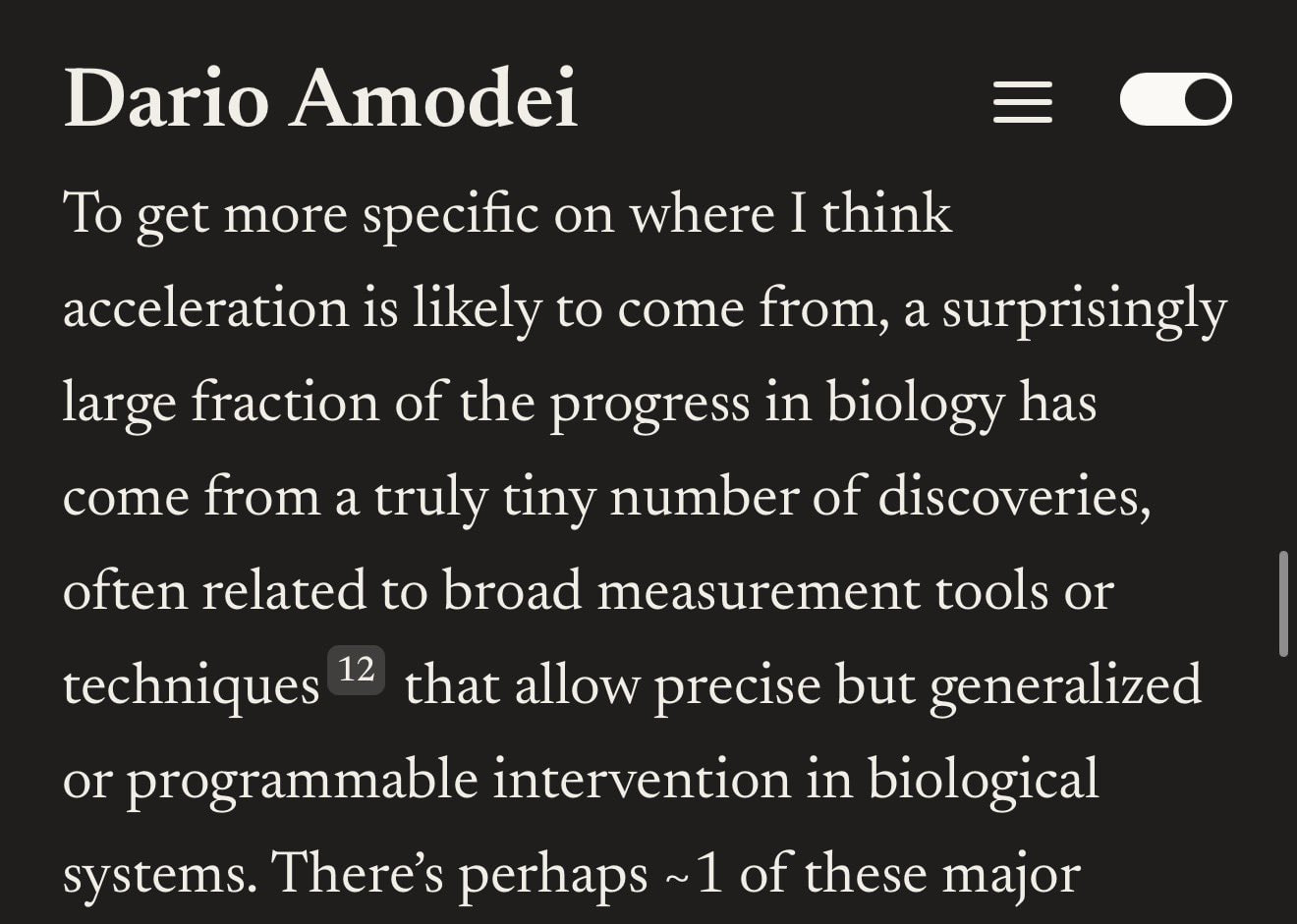
With AI doing the algorithmic side more and more, microscopic imaging improvements may now be one of the most important things
also cf Anshul Kundalje recent tweet
I remember he had a lot of lightfield microscopy: Research | Markov Biosciences
Together with UC Berkeley we are announcing the laser phase plate - a breakthrough in atomic resolution imaging. This is the brightest continuous wave laser in the world, 100 million times the intensity of the surface of the sun.
Phase contrast plays an important role in microscopy, but it was thought close to impossible for electron microscopy, where it would require interfering with an electron beam. Holger Mueller and Robert Glaeser proposed exactly this using a standing wave laser. It has taken over 15 years to make this a reality. Biohub partnered with UC Berkeley and Mueller to support this work and to engineer and build the technology.
Contrast has been the critical barrier to achieving atomic resolution imaging of the cell. In cryo-electron tomography, a cellular imaging technology that uses electron microscopy, the low contrast makes it impossible to resolve anything but the largest proteins within their cellular context. The laser phase plate removes that barrier.
With advances in AI this breakthrough in contrast will start to open up a new frontier in structural biology, that will allow us to see the molecular machines of the cell, and how they assemble into far more complex and dynamic systems, and understand how they work.
Nature biotechnology paper (15 July 26) reports a microscope that does something long considered impossible: RUSH3D-HR captures organelle-level detail across thousands of cells simultaneously in a living mammal, without killing them.
This builds on RUSH3D (Cell 2024) from the same group, which achieved single-cell resolution across a centimeter-scale surface. RUSH3D-HR narrows the imaging window roughly threefold to gain sevenfold lateral and ninefold axial resolution improvement, crossing from cell-tracking into organelle dynamics. The two instruments are designed as a pair.
Three experiments define the platform’s reach. In mouse skin wound healing, nearly 20,000 neutrophil trajectories over 400 minutes revealed organized net-like migration highways through adipose stroma before cells converge on wounds, invisible at smaller fields of view. In acetaminophen-induced liver failure, RUSH3D-HR caught a sixfold surge in migrasome generation within 50 minutes: these organelles nearly doubled their speed (0.13 to 0.29 µm/s) and over 70% gained new energy after forming versus under 20% in healthy mice, pointing to migrasomes as active signaling vehicles during liver injury. In inflamed mouse spleens, tracking over 6,000 neutrophils for five hours directly captured transient neutrophil swarms, coordinated aggregations hypothesized but never seen in living spleens. Electroacupuncture at the ST36 acupoint at 0.5 mA cut swarming by more than fivefold, the first direct cellular-scale evidence for how peripheral electrical stimulation dampens inflammation.
Limitation: The system only images 40 µm into tissue, limiting it to surface-accessible organs. Fluorescent labels are required. Only four color channels are available. The custom lens is not yet commercially available. The electroacupuncture result was demonstrated in a single inflammation model and needs broader validation.
Full text: https://lnkd.in/gwh7HFsA